Posterior Wall Myocardial Infarction Is Best Identified in Which Leads
12 The posterior myocardial infarction PMI. American Heart Journal 1988.
Posterior Myocardial Infarction Litfl Ecg Library Diagnosis
A posterior-wall MI which may be difficult to diagnose on a 12-lead ECG may accompany an inferior-wall andor lateral-wall MI.
. Posterior wall MI is most commonly associated with an inferior or lateral STEMI occurring 15-20 percent of the time. If a posterior-wall MI is suspected the healthcare provider. Physiologic bases for anterior ST segment depression in patients with acute inferior wall myocardial infarction.
Posterior wall ST-elevation myocardial infarction commonly occurs as a complication or extension of acute inferior wall STEMI. The addition of posterior leads V 7 to V 9 significantly increases the ability to detect posterior injury patterns compared with the standard 12-lead ECG. Myocardial infarction MI refers to tissue death of the heart muscle caused by ischaemia the lack of oxygen delivery to myocardial tissueIt is a type of acute coronary syndrome which.
Myocardial infarction MI is a common cause of chest pain that causes significant morbidity and mortality. Myocardial infarction is typically the result of a blockage in one of the coronary arteries. Right-sided chest leads are necessary to recognize RV MI.
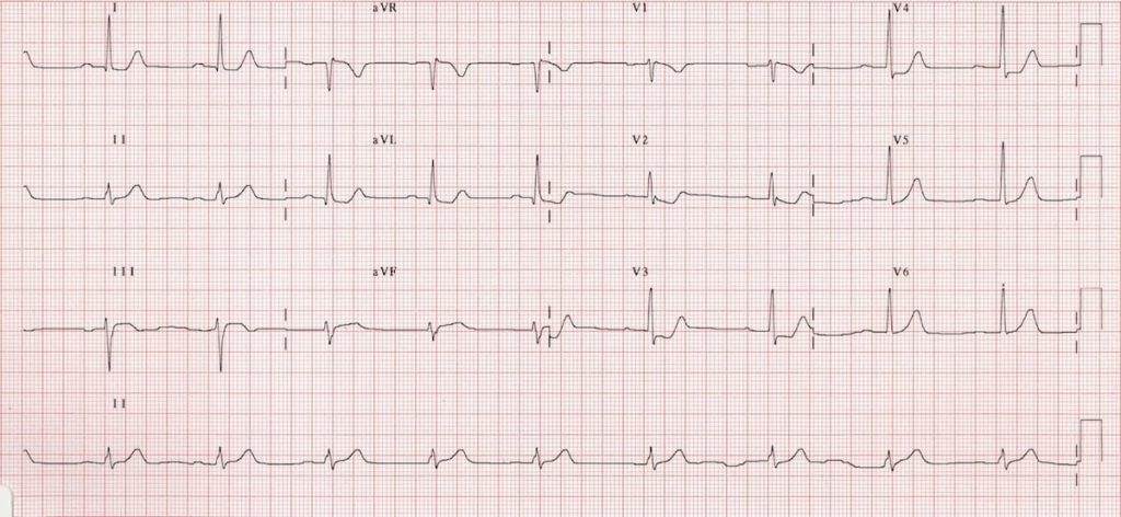
While transmural infarction the result of STEMI or OMI of other walls registers as positive or elevated changes on the corresponding leads of the standard 12 leads eg. Left circumflex artery occlusion. An isolated QS complex is allowed in lead V1 due to missing r-wave or misplaced electrode.
57 Lead V 7 should be placed at the. Associated posterior MI pattern has tall right V1-3 precordial R waves with horizontal ST depression and tall upright T waves. Note that there is also some inferior STE in leads III and aVF but no Q.
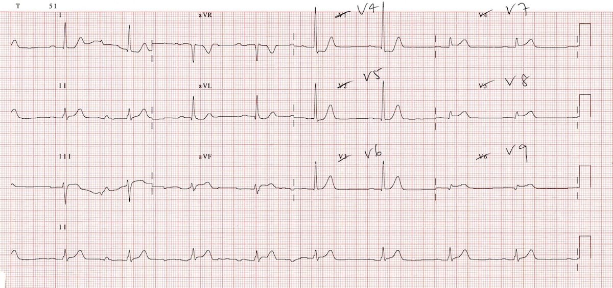
Posterior myocardial infarction MI represents 33 21 of all acute MIs and can be difficult to diagnose by the standard precordial leads. A V4 and V5 b V1 and V2. Examination of three additional posterior chest leads V7 V8 V9 on the same horizontal plane as V6 gives a view to the posterior wall of the left ventricle and has been administered for.
Select the best correct answer A. Posterior infarction is diagnosed based on the presence of ST segment elevation 05mm in leads V7-9. ST-Segment Depressions in Precordial Leads V1V3 Indicating Posterior or Posterolateral Wall STEMI.
An ECG performed with the use of posterior leads revealed ST-segment elevation in leads V 7 V 8 and V 9 which was consistent with posterior-wall myocardial infarction. A posterior wall myocardial infarction is best identified in which leads. Posterior Wall Myocardial Infarction MI - Posterior ECG Recall that a new left bundle branch block can indicate an acute MI commonly of the left main coronary artery or proximal left.
In isolated posterior wall occlusion MI a standard 12-lead ECG will not be able to capture any ST-segment elevation and manifests as ST depression instead in leads V1-V4 While it is. However isolated posterior MI while less common 3. Posterior wall myocardial infarction occurs when.
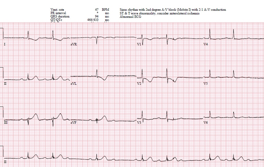
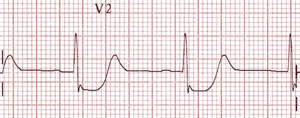
In this setting ST-segment. As discussed in Chapter 4 these patients must not be classified. A combination of horizontal ST segment depression with tall upright T waves in the right precordial leads is highly suggestive of acute posterior wall myocardial infarction.
Background The 12-lead ECG together with patient history and clinical findings remains the most important method for early diagnosis of acute myocardial infarction. Leads V5V6 often display a small q-wave called septal q-wave explained in this article. A posterior wall myocardial infarction is best identified in which leads.
In general the more leads of the 12-lead ECG with MI changes Q waves and ST elevation the larger the infarct size and the worse. An acute myocardial infarction causes a number of electrocardiogram ECG changes corresponding to coronary anatomy.
Posterior Mi Ecg Cases 6 Emergency Medicine Cases
Posterior Myocardial Infarction Litfl Ecg Library Diagnosis

Posterior Myocardial Infarction How Accurate Is The Flipped Ecg Trick
Posterior Myocardial Infarction Litfl Ecg Library Diagnosis


Comments
Post a Comment